The invention enable highly sensitive and selective fluorescent labelling of calcium deposits (e.g. hydroxyapatite) in tissue samples and allow the imaging detection of microcalcifications.
The molecules can be used in vitro and enable quantitative imaging of calcium deposits. However, they could also be used as fluorescent contrast agents in diagnostic imaging of calcifications in soft and hard tissues (e.g. vascular calcification, bone remodelling processes, microcalcifications in breast tissue, tissue ageing, macular degeneration).
Disturbances in calcium homeostasis can lead to abnormal tissue calcification, a feature of many diseases including macular degeneration, and certain cancers. Current clinical imaging techniques, such as radiography, fluoroscopy, CT, electron-beam tomography, intravascular ultrasound, and MRI, can detect larger calcifications but they lack the sensitivity to visualize microcalcifications below the millimeter scale. Many of these procedures may be invasive, costly, or poorly tolerated by patients. Fluorescent probes offer a promising alternative for highly sensitive, non-invasive detection; however, only a limited number of calcium-specific fluorophores currently exist.
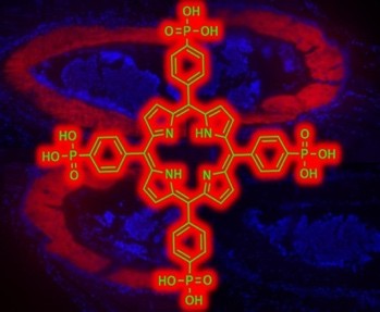
This patented technology introduces a new class of fluorescent probes specifically engineered to detect early-stage and micro-scale calcium deposits. These probes consist of an organic fluorescent core, such as porphyrins, phthalocyanines, acridines, BODIPY, or xanthene derivatives, which are functionalized with covalently bound phosphonate groups. These phosphonate groups bind calcifications with high affinity. Upon binding, the probes exhibit a distinct change in fluorescence intensity, enabling accurate visualization and quantification of calcified structures. Experiments with mouse tissue show clear fluorescent labelling of calcified structures. Initial tests on cell cultures confirm cell permeability and specific binding to calcium. The technology is at an advanced preclinical stage of development with promising results for diagnostic applications.
Ina Krüger
Technology Transfer Manager
+49 (0)30 314-75916
ina.krueger@tu-berlin.de
Technology demonstrated in relevant environment
pending: PCT, US
Technische Universität Berlin
The Center for Intellectual Property (ZfgE) at the TU Berlin is the central point of contact for all topics relating to intellectual property law and intellectual property.
We patent and market the inventions of the TU Berlin, and we also teach and research technical and intellectual property law.
This makes us the central contact for inventors of the TU Berlin, for cooperation partners from industry and science as well as for interested scientists and experts from the fields of technology and law.
Zentrum für geistiges Eigentum
Technische Universität Berlin
Straße des 17. Juni 135
10623 Berlin